
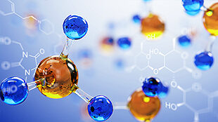
Control Ligands & Inhibitors
Highly purified, pre-validated reference ligands & inhibitors for use with Eurofins DiscoverX cell lines
Read More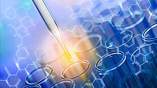
Detection Kits
Kit & reagents for sensitive quantification of biologics or small molecules to provide accurate pharmacology & reproducible results
Read More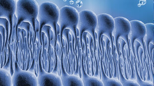
Membrane Preparations
Purified GPCR & ion channel membrane preps for binding & functional analysis
Read More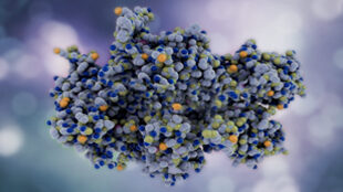
Recombinant Proteins
Over 500 high-quality & purified recombinant kinases, phosphatases, & epigenetic & ubiquitin proteins
Read More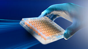
Assay Kits
Cell-based assay kits from early discovery to pre-clinical & clinical development to commercial release & stability programs
Read More
Toolbox Products
Complete set of parental cell lines, vectors, kits, & retroparticles to build your own stable cell lines & cell-based assays
Read More
Cell Lines & Primary Cells
Stable cell lines, cell pools, frozen cells, & primary effector cells for a broad range of target classes
Read More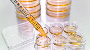
Cell Culture Kits & Reagents
Cell culture kits & reagents for cell culture preparation, maintenance of Eurofins DiscoverX cell lines, & running cell-based assays
Read More